Figure 1
From: Development of functionalised polyelectrolyte capsules using filamentous Escherichia coli cells

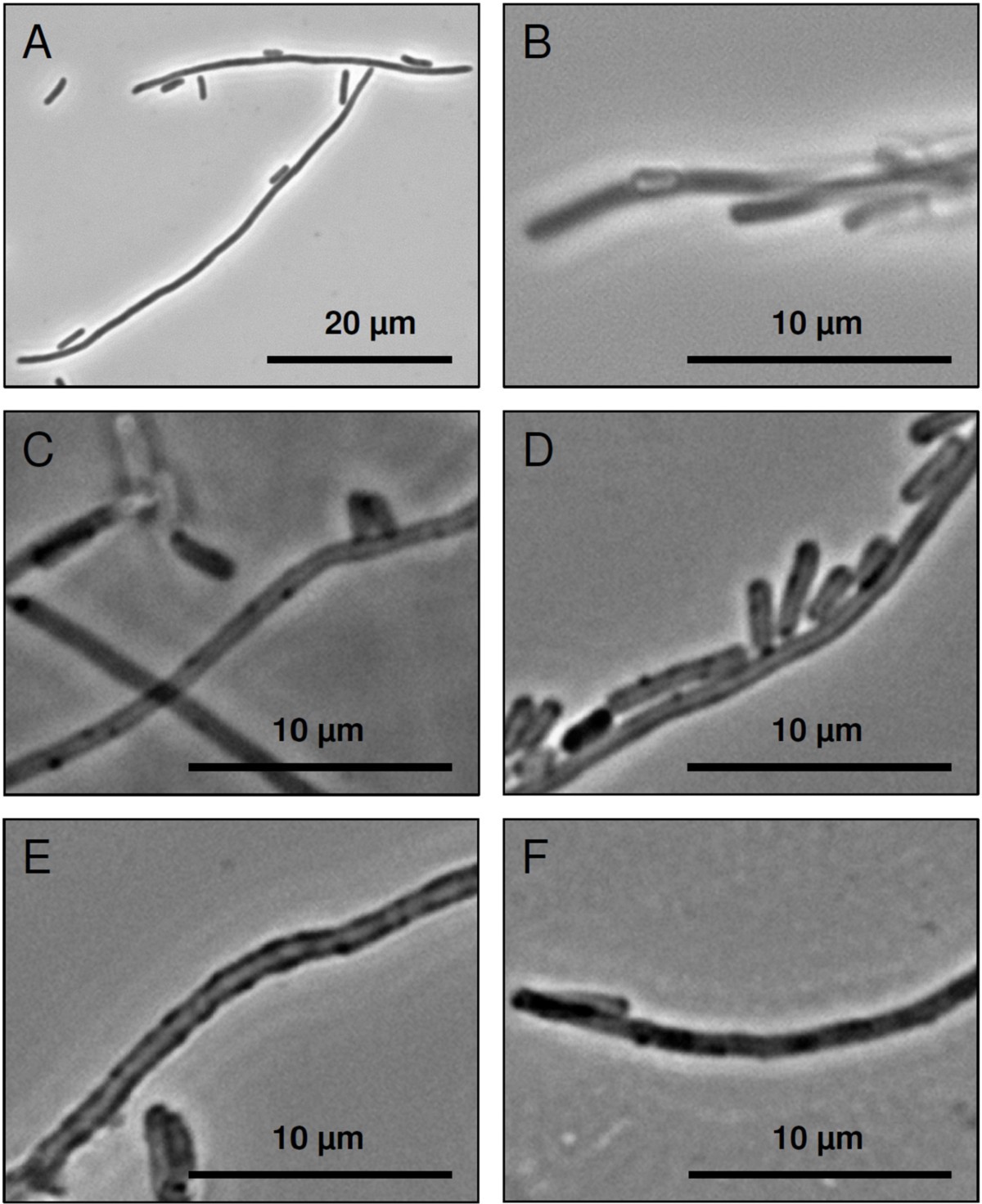
Light microscope images of filamentous E. coli cells and polyelectrolyte capsules in phase contrast mode. Image A presents filamentous E. coli cells in the exponential growth phase before polyelectrolyte coating. The polyelectrolyte coated E. coli filaments before NaOCl treatment are shown in image B. Image C presents polyelectrolyte tubes after the treatment with 1.2% NaOCl. The S-layer polymer protein coated polyelectrolyte capsules are shown in image D. Image E shows S-layer polymer protein coated polyelectrolyte tubes with synthesised palladium particles and image F presents polyelectrolyte capsules with synthesised palladium particles without S-layer proteins.